


Attention-Deficit/Hyperactivity Disorder is a prevalent neurodevelopmental disorder affecting millions globally, including an estimated 3%–7% of school-aged children, with symptoms often persisting into adulthood. Early and accurate diagnosis of ADHD…
Aging is a critical area of research because it is frequently accompanied by declines in sensory, motor, and attentional functions, which can significantly impair overall cognitive performance…
Understanding how we pay attention to the world around us is a fundamental question in cognitive science, particularly how our attention is captured…
In collaboration with the Department of English Language and Literature at Ferdowsi University of Mashhad, we proceed conducting several experiments in neurolinguistics mentioned briefly in the following…
Monitoring urine output is a critical diagnostic and management tool across various medical domains, including abdominal surgeries…
Emotions significantly influence human behavior, cognition, and decision-making. Automated emotion recognition systems are increasingly important for enhancing human-computer interaction…
Understanding how the human brain develops sensory capabilities before birth is a fundamental aspect of developmental neuroscience…
Sensory development of the human brain begins prenatally, allowing cortical auditory responses to be recorded at an early age in preterm infants…

© 2025 neurotracklab | All Rights Reserved
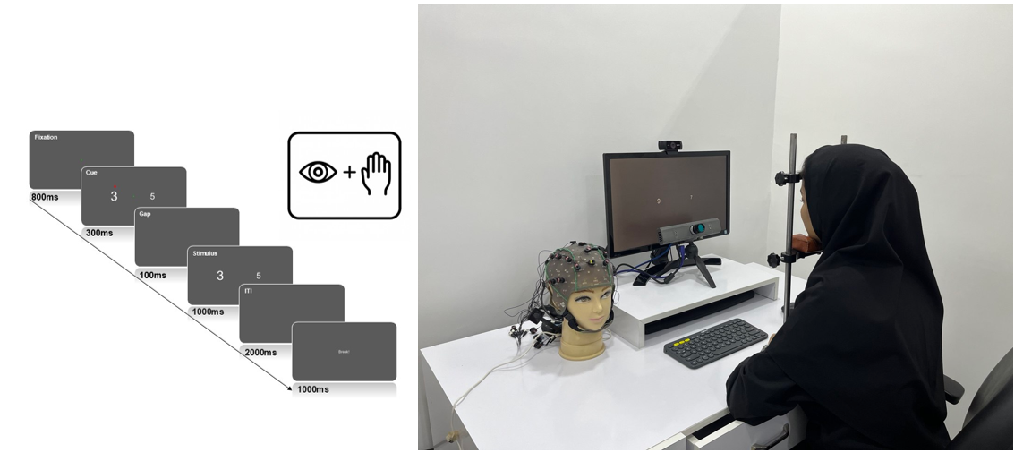
Attention-Deficit/Hyperactivity Disorder is a prevalent neurodevelopmental disorder affecting millions globally, including an estimated 3%–7% of school-aged children, with symptoms often persisting into adulthood. Early and accurate diagnosis of ADHD, particularly in adults, is crucial for timely and appropriate treatment, which can help prevent or reduce learning difficulties and social challenges.
Traditional ADHD diagnostic tools rely heavily on subjective evaluations, which are prone to bias and inconsistency. To overcome these limitations, this project proposes a novel multimodal approach for the objective diagnosis of ADHD in adults. Here, we combine behavioral performance analysis, eye-tracking, and galvanic skin response (GSR) measurements, integrated with supervised machine learning algorithms to detect ADHD. A customized version of the Stroop task is designed to simultaneously capture cognitive and physiological responses in controlled conditions. Eye movement features such as saccadic latency and fixation stability, alongside GSR-derived measures of autonomic arousal, serve as input features for predictive models. By fusing cognitive, physiological, and computational techniques, the study aims to build a robust framework for accurate ADHD detection and pave the way for personalized interventions. In collaboration with the Department of Psychiatry at Mashhad University of Medical Sciences, we aim to advance diagnostic approaches for neurological disorders so they can be broadly implemented in clinical practice.
Aging is a critical area of research because it is frequently accompanied by declines in sensory, motor, and attentional functions, which can significantly impair overall cognitive performance. Neuro-aging is dedicated to understanding the complex ways in which the brain changes as people grow older, focusing on the impact on cognitive function, sensory processing, and neural efficiency. These changes are commonly attributed to a reduction in the brain’s efficiency in processing stimuli, leading to reduced perception and a general decline in cognitive and motor abilities. Mechanisms such as diminished inhibition and impaired communication between brain regions, like the prefrontal cortex and thalamus, are often implicated.
Here, we focus on exploring how aging affects functional brain connectivity and the potential presence of compensatory mechanisms in the aging brain. By analyzing brain signals and event-related potentials (ERPs), the study examines different types of functional connectivity and investigates various computational methods for extracting connectivity features. The goal is to identify age-related changes in neural communication patterns and assess whether older adults recruit alternative networks to maintain cognitive function. Through this analysis, the project aims to contribute to a deeper understanding of neuroplasticity and functional adaptation in the aging brain.
Before proceeding to the functional connectivity analysis, in our previous work, we explored the correlations among microstructural, macrostructural, and functional aging-related changes in the human visual cortex, published in PLOS ONE and available online at https://doi.org/10.1371/journal.pone.0266206.
Understanding how we pay attention to the world around us is a fundamental question in cognitive science, particularly how our attention is captured by different visual stimuli. Visual information processing is profoundly shaped by attentional priority, which arises from both our current goals (goal-driven or endogenous attention) and the inherent properties of objects in the environment (salience-driven or exogenous attention).
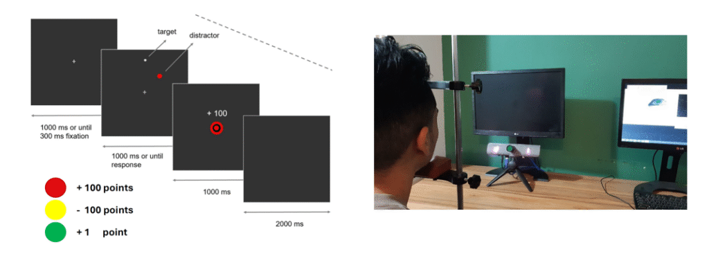
Recent research has highlighted a fascinating aspect of attention: its capture by stimuli that have previously been linked to rewards. This phenomenon, known as reward-driven attentional capture, demonstrates that even objects that are not physically prominent or are completely irrelevant to the task at hand can still grab our attention and guide our gaze, if they’ve been previously associated with a reward. For instance, studies have shown that if a particular color was previously associated to a high reward, a physically non-salient distractor with that same color could increase the time it took for participants to find a target in a subsequent task. Moreover, people tend to fixate more often on distractors signaling a high reward, and their reaction times to fixate on the actual target become slower in the presence of such distractors, irrespective of whether the distractor was physically salient. A critical question that remains, however, is whether this ‘learned value’ effect on attentional capture is driven by the motivational significance (salience) of the potential outcome, or by its valence (i.e., its positivity or negativity). In other words, is the effect of punishment identical to that of reward, or is it the opposite?
Here, we design and implement a series of experiments to investigate how reward- and punishment-associated visual stimuli influence attentional capture and oculomotor control. Using eye-tracking, we examine how stimuli linked to motivational outcomes affect the trajectory and dynamics of saccadic eye movements. In continuance to the above project, in our following researches, we aim to include the EEG analysis to seek if the reward and punishment (both motivationally significant) can further influence the EEG signals during the psychophysical tasks exploring the effect of valence and motivational salience on attention and target selection.

In collaboration with the Department of English Language and Literature at Ferdowsi University of Mashhad, we proceed conducting several experiments in neurolinguistics mentioned briefly in the following.
Given the association between multisensory information and inner attention, the quality of multisensory information is hypothesised to modulate the neuronal activity associated with language comprehension. To verify, we investigated the interaction effect between multisensory quality (sensory enrichment) and semantic (in)congruency during L2 sentence comprehension. The reader may refer to “Oscillatory neuronal dynamics during L2 sentence comprehension: the effects of sensory enrichment and semantic incongruency” for the details.
We also examined the role of the combination of three senses (i.e., auditory, visual, and tactile) and five senses (i.e., auditory, visual, tactile, olfactory, and gustatory) in the correlation between electrophysiological and electrodermal responses underlying second language (L2) sentence comprehension. The reader may refer to “Cognition-Emotion Interaction during L2 Sentence Comprehension: The Correlation of ERP and GSR Responses to Sense Combinations” for the details.
Our latest research is related to demonstrating a link between specific brainwave frequencies and various linguistic abilities. It is postulated that observable associations will exist between an individual’s resting-state brainwave patterns and their overall language proficiency. To the best of our knowledge, the correlation between L2 proficiency and neural signatures in open-eyes and closed-eyes resting state has not been extensively examined. In this study, we use EEG to benefit from high-quality temporal resolution data. While resting-state EEG provides valuable insights into neural activity, it is important to note that it does not directly assess language processing. Given its sensitivity to various cognitive and neural factors, caution should be exercised when linking observed differences to language proficiency.
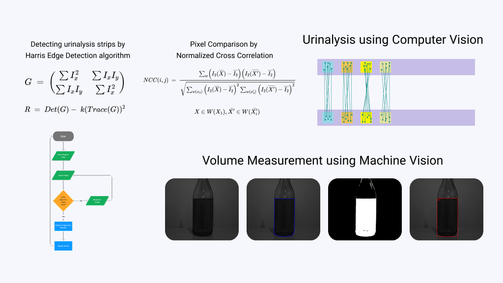
Monitoring urine output is a critical diagnostic and management tool across various medical domains, including abdominal surgeries, high-risk pregnancies, severe burns, cardiovascular diseases, acute kidney injury (AKI), sepsis, cancer, prostate disorders, and spinal cord injuries. It plays an essential role in the early detection of complications and the optimization of clinical outcomes. Currently, urine output measurement in hospital settings relies predominantly on manual methods, utilizing graduated urine collection bags inspected by nursing staff at approximately hourly intervals. This approach lacks precision and fails to capture real-time fluctuations in a patient’s clinical status. The rising incidence of conditions such as diabetes, alcohol dependency, and renal diseases further underscores the urgent need for precise and continuous urine output monitoring to mitigate complications such as oliguria, hematuria, and disease metastasis.
To address this critical need, we propose the design and development of an advanced, intelligent system for real-time measurement and analysis of patients’ urine output. This system integrates multiple subsystems, including urine inflow detection, precise volume measurement, inter-chamber transfer, qualitative parameter analysis, and data transmission for automated prognosis and screening of critical medical conditions. The measurement of urine volume and qualitative analysis are facilitated by machine vision technology, leveraging sophisticated image processing algorithms to ensure high accuracy in identifying urine characteristics. Additionally, the system incorporates a machine learning module specifically tailored for the detection and prediction of AKI, enabling the identification of relevant patterns through comprehensive analysis of input data. The system is further equipped with an audio-visual alert mechanism that promptly notifies healthcare providers of anomalies, such as abnormal reductions in urine volume or qualitative changes indicative of AKI. Designed to be universally applicable across genders, this system is versatile for use in diverse medical scenarios, including surgeries, burn care, high-risk pregnancies, and spinal cord injury management.
Emotions significantly influence human behavior, cognition, and decision-making. Automated emotion recognition systems are increasingly important for enhancing human-computer interaction, improving mental health monitoring, and developing more empathetic assistive technologies.
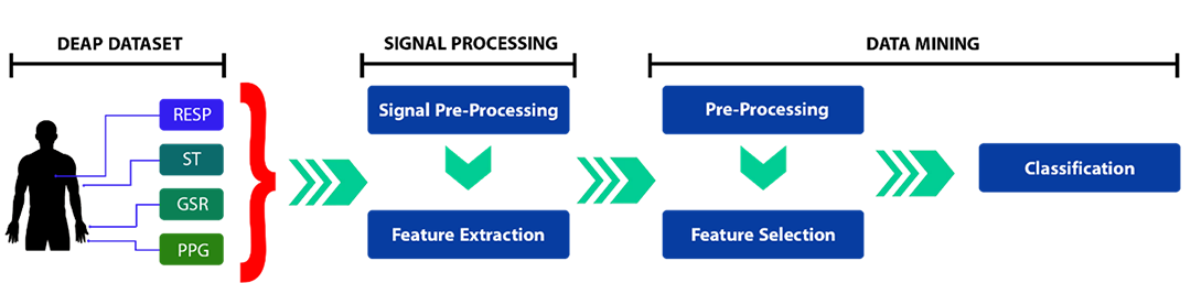
Physiological signals offer a robust and objective means for emotion detection, as they capture involuntary bodily responses to emotional stimuli. Signals such as Photoplethysmography (PPG), Respiration Rate (RR), Skin Temperature (ST), and Galvanic Skin Response (GSR) are particularly valuable due to their connection with the autonomic nervous system. By integrating multiple physiological modalities, we can overcome the limitations of individual sensors and achieve more accurate and resilient emotion recognition.
Our work introduces a machine learning-based approach to classify emotions within a two-dimensional valence-arousal model (pleasantness and intensity of emotion). We extract comprehensive features from multiple physiological signals and employ advanced feature selection techniques to identify the most informative data points. This systematic methodology contributes to the development of real-time emotion recognition systems by optimizing signal processing, feature selection, and classification performance, providing a robust framework for future advancements in affective computing.

Understanding how the human brain develops sensory capabilities before birth is a fundamental aspect of developmental neuroscience. Functional responses recorded during the last trimester of gestation reveal that sensory activity begins before birth, allowing the brain to process the external environment even in preterm infants. As the brain matures, new cognitive skills emerge, and studying the intricate relationship between this structural maturation and functional development in vivo is crucial for characterizing normal brain development and understanding the early mechanisms of neurological conditions.
Here, we aimed to utilize non-invasive techniques to investigate the relationship between the brain structure and function by relating developmental changes in cortical auditory evoked potentials (CAEPs) to a structural maturation index, primarily representative of myelination. We recorded CAEPs in preterm neonates in response to auditory stimuli and extracted specific functional features, such as the latency of the global field power (GFP). To assess structural maturation, we used T1-weighted (T1w) and T2-weighted (T2w) MRI images to derive maturation indices for various brain regions, including those involved in auditory processing. This allowed us to develop conjoint models of functional and structural brain development in this critical early period of life. https://doi.org/10.1007/s00429-020-02117-3
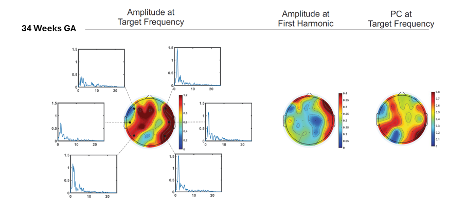
Sensory development of the human brain begins prenatally, allowing cortical auditory responses to be recorded at an early age in preterm infants. Despite several studies focusing on the temporal characteristics of preterm infants’ cortical responses, there’s a recognized gap in understanding their frequency-domain properties.
Our study addressed this gap by performing frequency and coherence analysis of preterm infants’ auditory responses to repetitive syllabic stimuli. We also delved into the functional brain asymmetry of preterm infants, examining how their brains detect the regularity of auditory inputs. Our methodology involved recording cortical auditory evoked potentials (CAEPs) and extracting specific features like peak amplitudes of frequency responses and phase coherence at target frequencies. Additionally, we defined a functional asymmetry coefficient to assess lateralization, contributing to a deeper understanding of hemispheric specialization in the developing brain. Our findings showed for the first time that the right hemisphere lateralization for slow rate modulation, previously observed in adults, children and newborns, appears to be in place at a very young age, even in preterm infants (published by Nature Portfolio in Scientific Reports, https://doi.org/10.1038/s41598-019-47064-0).